Case

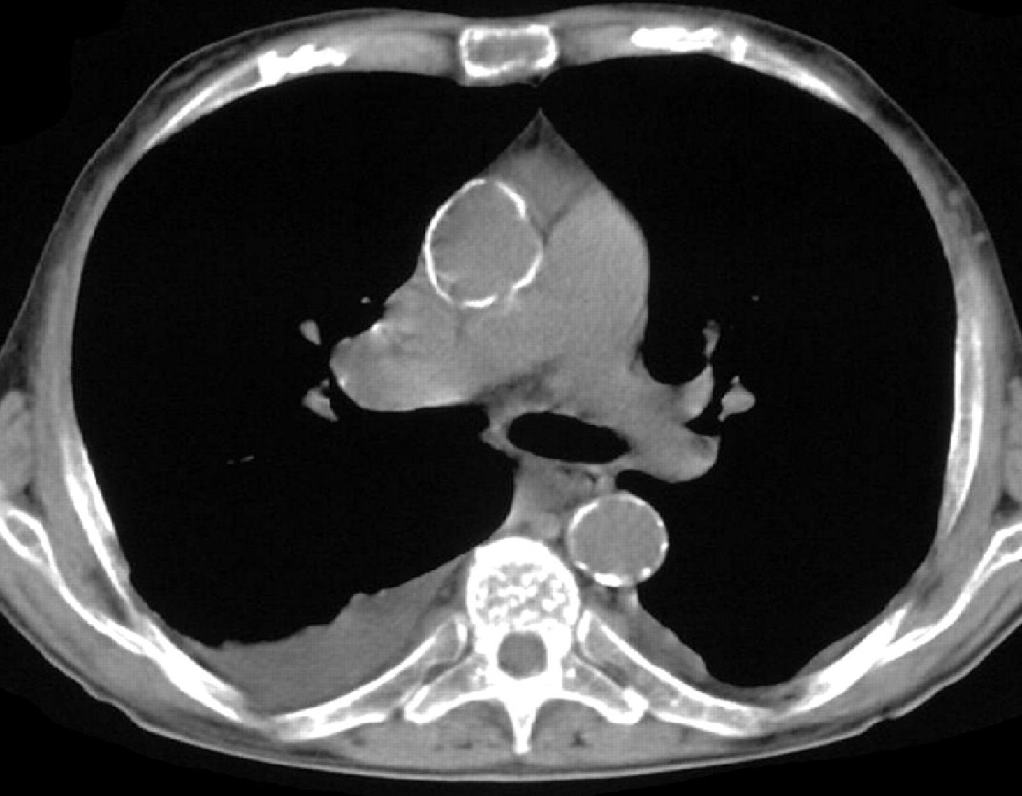
An 83 year old patient comes to the pulmonology division complaining of shortness of breath with any physical activity, persistent fatigue, and periods through the day where they reports having rapid and somewhat irregular heartbeat. Physical examination is otherwise unremarkable. A CT scan is ordered.


Question 1/3 - Approximately what level of the thorax do you think this CT is from?

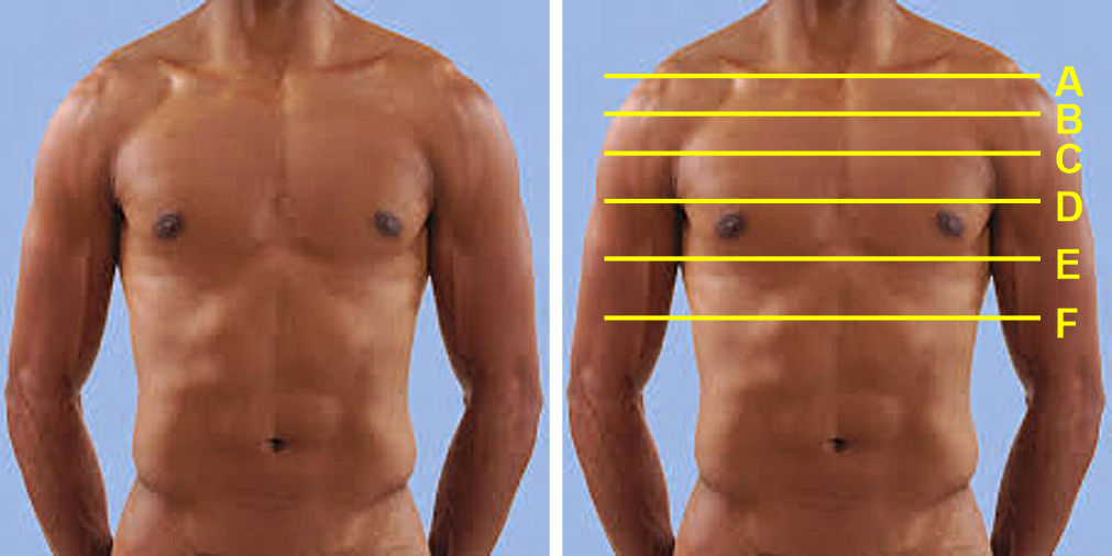
Click on your selected option(s) below  (correct = 1, over-thinking = 2+)

Incorrect. This line runs through the clavicles, which are not present on the CT scan.

Incorrect. This line is approximately at the level of the sternoclavicular joint, which we cannot see any clavicle in the CT scan

Correct!  The presence of the scapula posteriorly indicates the scan is from the upper chest. The absence of the clavicles indicates the level is inferior to the sternoclavicular joint.

Soft tissues show two aorta cross-sections and the absence of the heart. This puts the level approximately near the start and end of the aortic arch. Recall, the start and end of the aortic arch is a landmark for the sternal angle. Thus, the level of this CT is several inches inferior to the clavicles right around the sternal junction of the 2nd ribs (i.e. sternal angle).

We also see a dark region elongated as an oval indicating the trachea starting to bifurcate to the left and right bronchi (thus forming an 'oval' as it starts to branch to each side). That is another key feature of the sternal angle landmark.

Incorrect, but close.  A significant amount of scapula is present in the CT image, whereas the D level is probably only at the inferior tip of the scapula.  Also, as D is mid-thorax we would expect heart chamgers to be present on the scan, however all that is visible in the CT are the great vessels indicating the image is a level just above the heart.

Incorrect. This level is close to the xiphoid process and well below the level of the scapula bones which are visible in the CT scan.  Also, this low in the thorax heart chambers should be visible, while in the image they are absent.

Incorrect.  This level is inferior to the xiphoid process so no sternum or scapula would be present, but both are in the image indicating F is incorrect. Furthermore, F is sufficiently inferior that stomach and abdominal contents would be expected.